
25 February 2025
Future Healthcare Innovations
in Medical Treatment

Digital X-rays
Digital X-Rays are an advanced imaging technology used to capture detailed images of the internal structures of the body quickly and accurately. Unlike conventional film-based radiography, digital systems use electronic sensors to produce high-resolution images that can be instantly viewed, enhanced, and stored. This modern approach allows for faster diagnosis while minimizing radiation exposure and improving overall patient safety.
At Hridaydeep Heart Clinic & Diagnostic Center, Digital X-Ray examinations are performed using state-of-the-art equipment to ensure clarity, precision, and efficiency. The procedure is simple, painless, and typically completed within minutes, making it suitable for evaluating a wide range of conditions, including bone injuries, chest and lung concerns, and structural abnormalities. The ability to digitally process and share images enables doctors to assess results promptly and plan appropriate treatment without delay. By combining speed, accuracy, and reduced radiation exposure, Digital X-Rays play a vital role in comprehensive diagnostic care, supporting early detection and effective medical decision-making. By offering accurate data that guides personalized care planning, Laboratory and Pathology Services contribute significantly to proactive health management and improved long-term wellness.
OPG
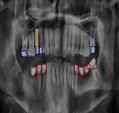
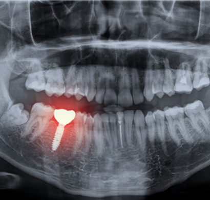
Orthopantomogram, commonly known as OPG, is a specialized panoramic dental X-ray that provides a comprehensive view of the entire mouth in a single image. It captures detailed visuals of the upper and lower jaws, teeth, jaw joints, sinuses, and surrounding bone structures, making it an essential diagnostic tool in modern dental and maxillofacial care. Unlike traditional intraoral X-rays that focus on a small area, OPG offers a wide-angle image that helps dentists and specialists evaluate overall dental health efficiently. It is widely used for detecting impacted teeth, wisdom teeth positioning, cavities, bone loss, jaw fractures, infections, cysts, tumors, and other abnormalities affecting oral and facial structures.
The OPG scan is quick, painless, and non-invasive. During the procedure, the patient stands or sits still while the machine rotates around the head, capturing a full panoramic image within seconds. The radiation exposure is minimal, making it a safe option for routine dental assessments and treatment planning. OPG plays a crucial role in various dental procedures, including orthodontic planning (braces), dental implants, root canal treatments, and oral surgeries.
CBCT
Cone Beam Computed Tomography (CBCT) is an advanced and highly precise 3D imaging technology used to visualize detailed structures of the teeth, jaw, face, and surrounding areas. It provides high-resolution, three-dimensional images that allow doctors to accurately assess bone structure, soft tissues, nerves, and airways, making it an essential tool in modern diagnostic and treatment planning.
Unlike traditional X-rays, CBCT captures multiple images from different angles and reconstructs them into a comprehensive 3D view. This enables better evaluation of complex anatomical structures that cannot be clearly seen in standard 2D imaging. CBCT is widely used in dental, maxillofacial, and ENT applications, helping in procedures such as dental implant planning, orthodontic assessment, root canal evaluation, jaw disorders, sinus analysis, and trauma cases. One of the key advantages of CBCT is its ability to provide highly accurate and detailed images with relatively lower radiation exposure compared to conventional CT scans. The scan is quick, painless, and typically completed within a few minutes. During the procedure, the patient simply stands or sits still while the machine rotates around the head, capturing precise images without discomfort.
HSG
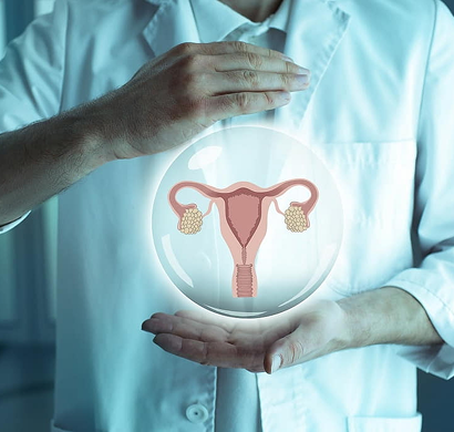
Hysterosalpingography, commonly known as HSG, is a specialized diagnostic procedure used to evaluate the uterus and fallopian tubes, primarily in women experiencing infertility or reproductive health concerns. This test uses a safe contrast dye along with X-ray imaging to provide clear and detailed visualization of the uterine cavity and the patency (openness) of the fallopian tubes. HSG plays a crucial role in identifying conditions that may affect a woman’s ability to conceive. It helps detect blockages in the fallopian tubes, abnormalities in the shape or structure of the uterus, adhesions (scar tissue), fibroids, polyps, or congenital defects.
The procedure is usually performed within a few days after the menstrual cycle ends to ensure accuracy and safety. During the test, a thin catheter is gently inserted into the cervix, and a contrast dye is introduced into the uterus. As the dye flows through the fallopian tubes, X-ray images are taken to track its movement and identify any blockages or irregularities. HSG is a relatively quick procedure, typically completed within 15 to 30 minutes. While some patients may experience mild discomfort or cramping during the test, it is generally well-tolerated and does not require hospitalization. The results are available shortly after the procedure, allowing doctors to recommend further steps such as medication, minimally invasive procedures, or assisted reproductive techniques if needed.